


Screening & Diagnosis Solution
Time is precious when it comes to effective screening, detection and diagnosis of breast cancer. We strive to improve efficiencies at every step, ensuring more women have more time in better health.
Advance Early Detection & Diagnostic Accuracy
Our clinically proven, integrated breast imaging and diagnostic solutions can optimise your clinical pathway while enhancing a woman’s breast imaging experience.
Cutting-Edge Imaging
Optimal cancer detection starts with the highest quality imaging. Integrate the EUREF-approved1 Hologic mammography technologies into your clinical pathway to maximise efficiency in your imaging workflow.
Confident Cancer Detection
The clinically proven tomosynthesis and contrast enhanced imaging technologies, plus future-proofed AI powered solutions at the point of care, help you make the best clinical decisions while accelerating reading time and improving operational efficiency.
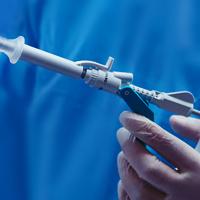
Compassionate Interventional Solutions
Bringing high quality imaging and intervention together helps you deliver more compassionate and effective solutions. Choose from stereotactic, ultrasound and MRI compatible biopsy markers. Tailor biopsy treatment options in prone or upright mode to help improve the patient experience.
Diagnostic & Data Efficiency
Powerful high-resolution imaging solutions require integrated, streamlined and breast-specific workflows. Our diagnostic workstations, scalable archiving and versatile image routing solutions can optimise reading workflow, minimise network burden and grow with your clinical needs.
Decades of Continuous Innovation
Our pioneering technology supports best-in-class detection and diagnostics,* delivered with speed and accuracy.1
3.7s
Scan Time, the industry’s fastest tomosynthesis scan² on the 3Dimensions™ Digital Mammography System**
1h
Daily saving in image interpretation time3,4 with 3DQuorum™ Imaging Technology
70 Micron
sharper images with Clarity HDTM High Resolution Tomosynthesis5
Browse our Flagship Range
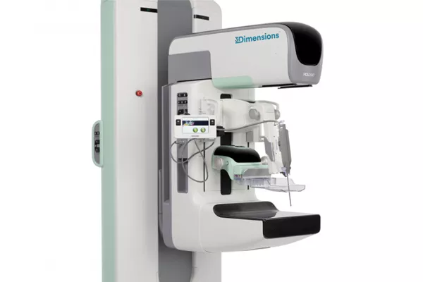
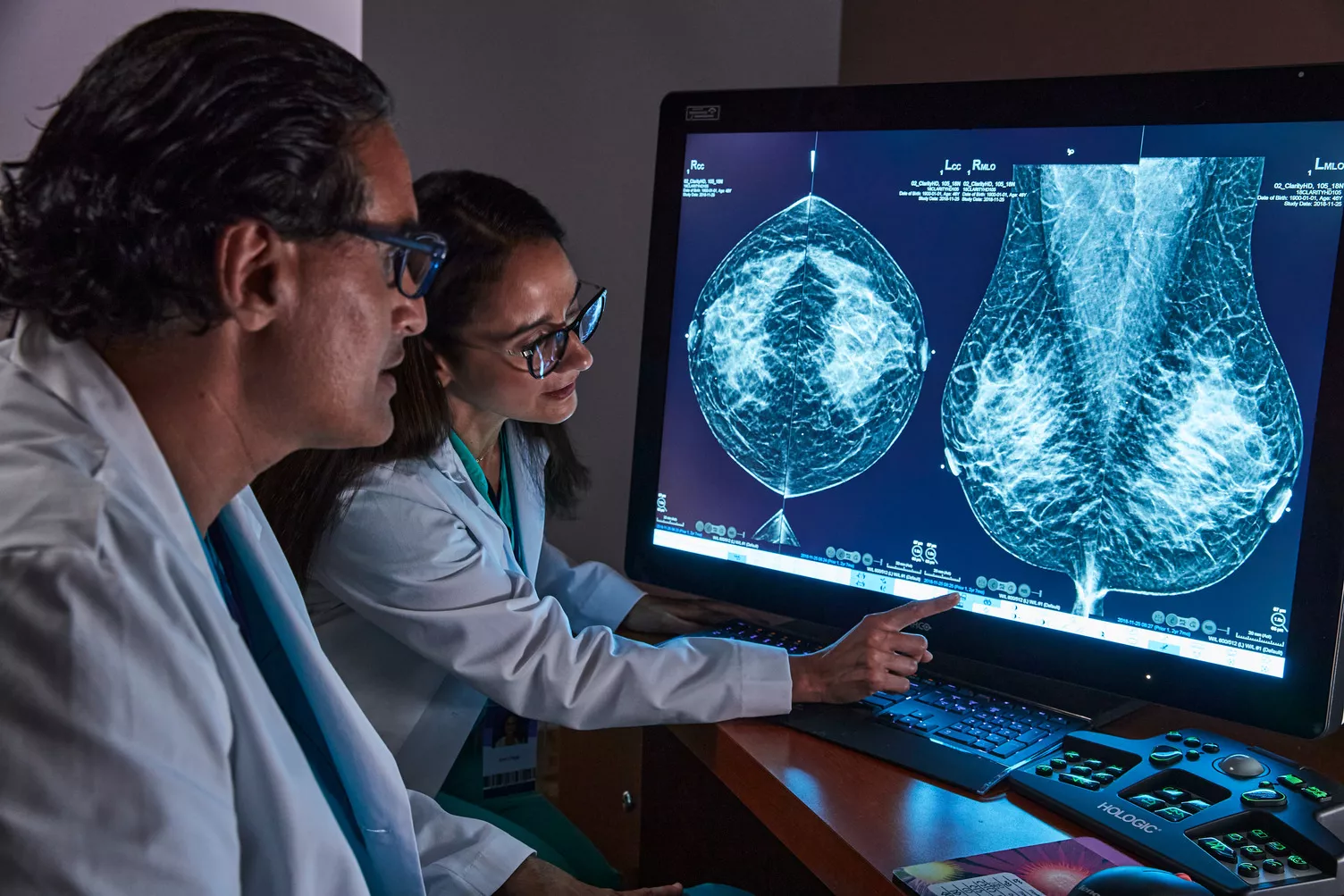
3Dimensions™ Digital Mammography System
Reveal fine details with the fastest, highest resolution 3D Mammography™ exam5 using our advanced detector and innovative 3D™ imaging technologies. These help to detect more invasive cancers with confidence6 With the integration of additional AI-powered solutions, this is a smart platform supporting breast cancer detection, workflow optimisation and risk assessment.
Clarity HD™ High Resolution Tomosynthesis
Producing high-resolution tomosynthesis images normally means slower read times. However, combining Clarity HD high-resolution 3D imaging with Genius AI Detection technology and 3DQuorum allows you to unleash the fastest and highest resolution 3D images in the industry,5 accelerating screening and analysis.
Intelligent 2D™ Synthesised Imaging
For higher image quality, more detail, improved read times and low dose, this AI-powered software produces robust, yet natural looking, synthesised 2D images that are well correlated with the 3D mammography data. This enables radiologists to quickly and confidently see subtle mammography features and lesion morphology.
3DQuorum™ Imaging Technology
3DQuorum technology utilises Genius AI powered analytics to uniquely reconstruct high-resolution 3D Mammography data to produce 6mm SmartSlices. It speeds up reading time by reducing the number of images to review, with no compromise in image quality, sensitivity or accuracy.7,8
Genius AI™ Detection Technology
A deep-learning algorithm designed to aid radiologists' diagnostic performance and detect breast cancer4,9,10 from tomosynthesis images from the Hologic Dimensions Mammography Systems. It locates lesions by searching each slice of the image set. Suspicious areas are highlighted for concurrent reading at the workstation.
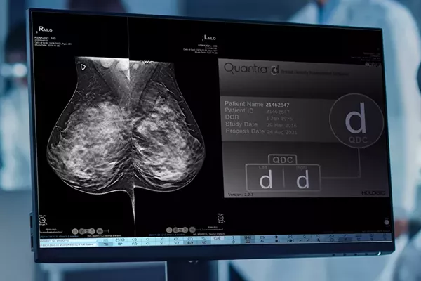
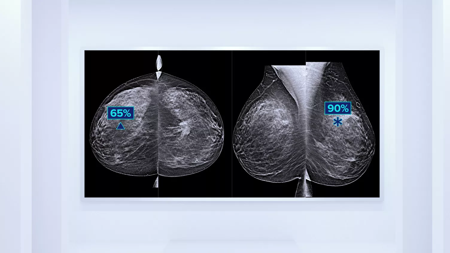
Quantra™ 2.2 Technology
Higher breast density is known to increase a woman’s risk for breast cancer.11,12 The need for accurate, unbiased analysis is therefore critical. Powered by machine learning, Quantra software analyses the breast tissue texture and pattern in both 2D and tomosynthesis images and categorises the breasts into four composition risk categories.13
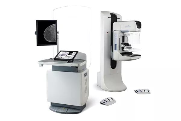
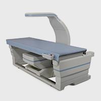
Selenia® Dimensions® Digital Mammography System
The Selenia Dimensions system delivers proven accuracy of our 3D Mammography exam to detect significantly more invasive breast cancers earlier and reduce call backs vs 2D alone.14-16† The system comes in two main configurations, to which you can add a range of options.
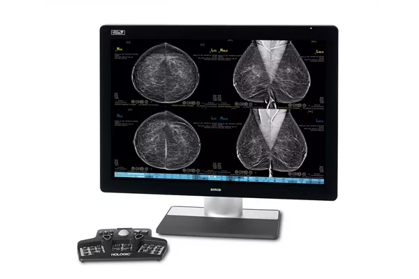
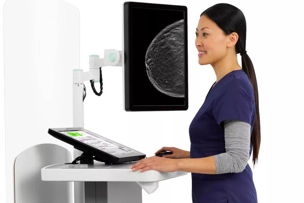
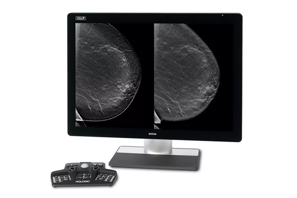
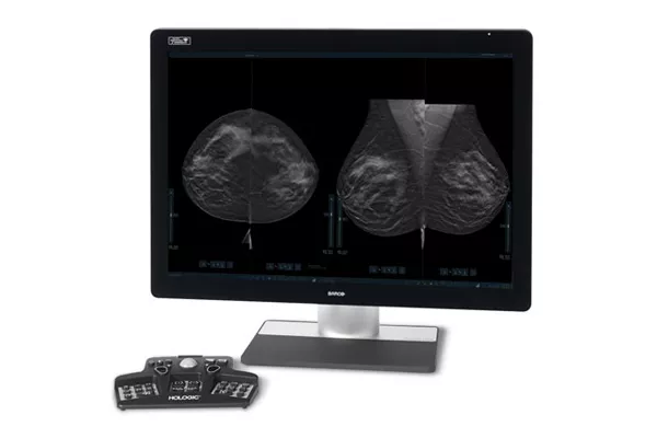
SecurView® Breast Imaging Workstation
A breast diagnostic workstation designed by radiologists for radiologists. Speed up your diagnosis with customisable workflow tools to ensure accurate, efficient evaluation of 2D and 3D Mammography exams. It does this through fast, configurable scrolling, on-the-fly slabbing, focus and co-registered 2D and 3D mammography exam images.

Unifi™ Connect
Unifi Connect provides a secure infrastructure for a full range of remote services for Hologic instrumentation, creating a continuous link between our technical experts and the customer. We monitor, identify and address issues remotely, maximising uptime and reducing the need for onsite visits by our engineers.
Insights


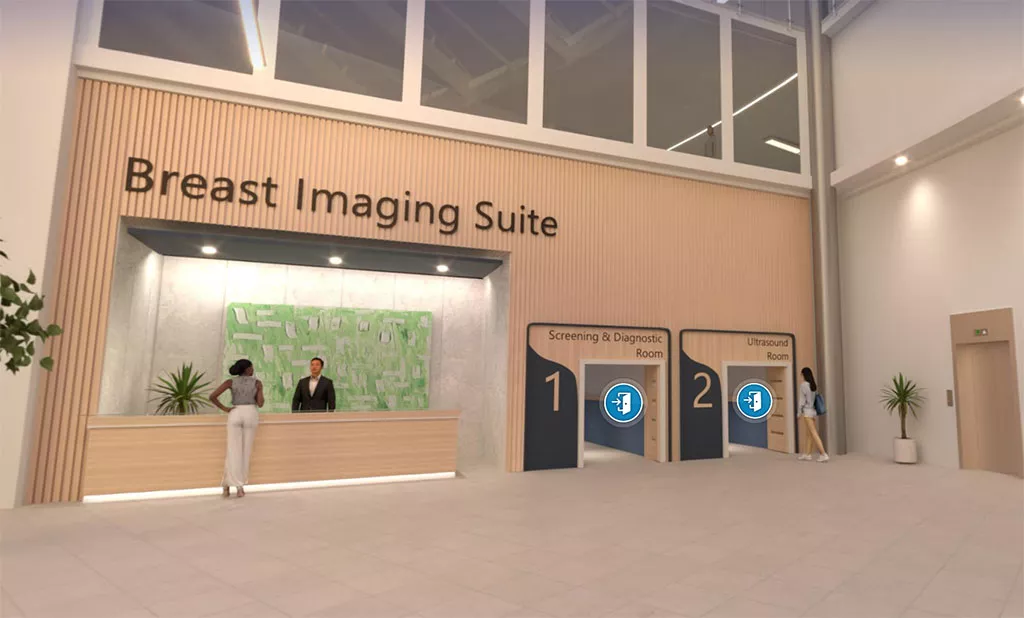
Visit Our Virtual Hospital
Browse our portfolio of Breast Health solutions in 3D. See how you can unlock the advantage of time across the entire Breast Continuum of Care.
* The only system to have received EUREF certification for both 2D and 3D.
** Compared to other standard models.
† When using 3D plus 2D exam
- Results EUREF type test. Available at: https://www.euref.org/type-test-equipment/euref-type-test-results (accessed November 2022)
- Rocha Garcia, A.M., Mera Fernandez, D. Breast tomosynthesis: State of the art. Radiologia. 2019;61(4):274-285.
- Hologic data on file: CSR-00130 Rev 001
- Data on File: Clinical Study Report CSR-00116, Rev 004
- Compared to other standard models. Rocha Garcia, A.M., Mera Fernandez, D. Breast tomosynthesis: State of the art. Radiologia. 2019;61(4):274-285
- Friedewald SM., Rafferty EA., Rose SL., et al. Breast cancer screening using tomosynthesis in combination with digital mammography. JAMA. 2014 Jun 25;311(24):2499-507.
- Hologic data on file: MAN-06153 Rev 002. 3DQuorum Physician’s Labeling
- Hologic data on file: MAN-06029-002 Rev 004 3D Quorum User Guide
- Hologic data on file: TFS-00002 Rev 003.
- Hologic data on File: DHM-09325 Rev 003.
- Rafferty EA, Durand MA, Conant EF, et al. Breast Cancer Screening Using Tomosynthesis and Digital Mammography in Dense and Non-dense Breasts. JAMA. 2016 Apr 26;315(16):1784-6.
- Dense breast higher risk factor. Available at: https://www.cancer.gov/types/breast/breast-changes/dense-breasts#:~:text=Dense%20breasts%20are%20not%20considered,than%20women%20with%20fatty%20breasts. (Accessed Nov 2022)
- ACR BI-RADS Atlas 5th Edition. 2013. Available at: https://www.acr.org/Clinical-Resources/Reporting-and-Data-Systems/Bi-Rads (Accessed Nov 2022).
- Tozaki M, Fukuma E. Pattern classification of ShearWave™ Elastography images for differential diagnosis between benign and malignant solid breast masses. Acta Radiol. 2011;52(10):1069-1075. doi: 10.1258/ar.2011.110276.
- Rose SL, Tidwell AL., Bujnoch LJ., et al. Implementation of breast tomosynthesis in a routine screening practice: an observational study. AJR AmJ Roentgenol. 2013;200(6):1401-1408
- Haas BM., Kalra V., Geisel J., et al. Comparison of tomosynthesis plus digital mammography and digital mammography alone for breast cancer screening. Radiology 2013;269:694-700.
Related Portfolio & Solutions
Breast Health Continuum of Care
Time is precious when it comes to effective detection, diagnosis and treatment of breast cancer. We strive to save you time at every step along the Breast Health Continuum of Care ensuring more women have more time in better health.
2797
Hologic BV, DA Vincilaan 5, 1930 Zaventem, Belgium
Notified Body number wherever applicable
EC Representative Information wherever applicable