

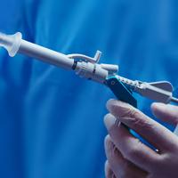


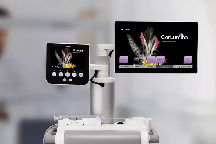
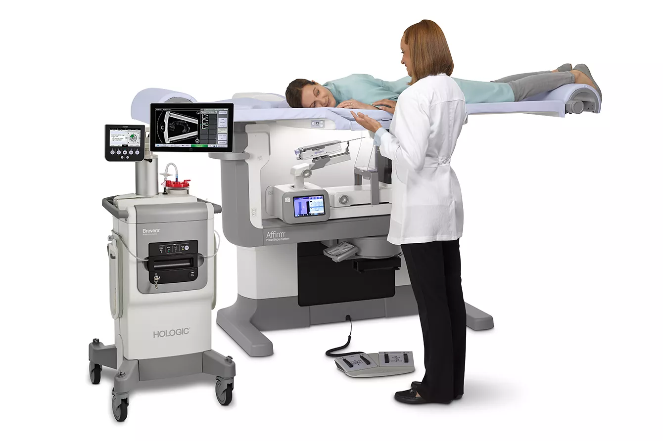
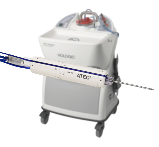
Brevera® Breast Biopsy System
Real-time imaging for instant sample verification during breast biopsy procedures.1-3
Update on Biopsy Needle Availability
Hologic is committed to the highest quality and is working urgently to address market shortages following a field safety notice and the removal of all Brevera® Breast Biopsy System 9 Gauge needles in December 2025. We are currently targeting return of the Brevera Breast Biopsy 9 Gauge needles to market by the end of the calendar year 2026.
In the interim, we are committed to supporting your operations through the following immediate actions:
- Increasing Biopsy Needle Supply: We are significantly increasing the supply of Eviva® needles. This is backed by major investments in our internal manufacturing, including additional personnel resources and expanded manufacturing shifts.
- Expediting Product Delivery: We are expediting shipping on all orders to ensure you receive essential supplies as quickly as possible.
- Stabilising Inventory: Our teams are working closely with component suppliers to increase volumes and restore long-term inventory consistency.
Eviva needles continue to be allocated through a multi pronged approach that considers historical ordering patterns prior to the field action, procedure volume and inventory levels to support planning.
Throughout this process, we are working closely with local country competent authorities in communicating ongoing activities and return to market for the needles. We remain committed to providing regular updates here as they become available. Customers may contact their Hologic sales representative with any questions.
Gain the Advantage of Time
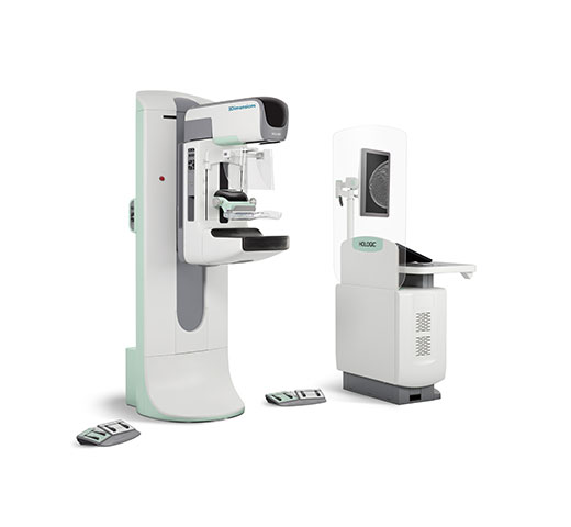
We understand that reducing time under compression for your patients during a breast biopsy procedure is essential to deliver an improved patient experience. The Brevera Breast Biopsy system in combination with CorLumina® Imaging Technology streamlines the entire breast biopsy process from start to finish. This includes real-time imaging for instant verification and automated post-biopsy specimen handling.1-3
Reduced Procedural Steps
There is no need to move to another room for imaging, no waiting for sample verification and fewer interruptions to your screening schedule. Instead, you can experience the detailed, real-time, accurate information you need to improve patient care and enhance your clinical workflow.1-3
Improve Patient Experience
Fast, accurate and streamlined procedures mean less time under compression and result in a more positive and compassionate biopsy experience for you and your patient.2,3
Enhance Workflow
An intuitive user interface, real-time imaging, and automated specimen collection and separation work together to save facilities an average of 13 minutes per procedure.2,3
Increase Accuracy
Real-time imaging delivers a wealth of information at the point of care – so you can make informed clinical decisions with confidence.1-3
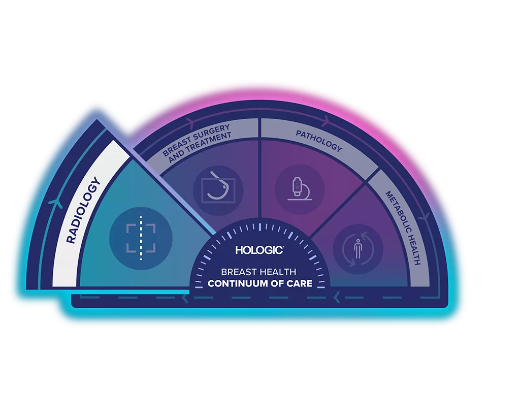
Unlock the Advantage of Time
The Breast Health Continuum of Care offers integrated solutions for clinical confidence, workflow efficiency and compassionate patient care. It gives more women, more time in better health.
The Brevera Breast Biopsy System is part of the Hologic Biopsy Solution.
Key Features & Benefits
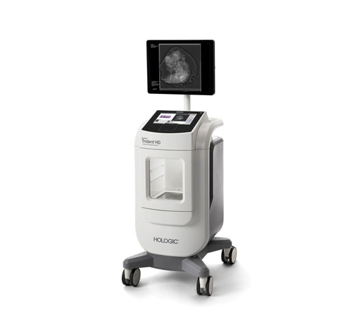
- Instant tissue verification with CorLumina imaging technology1
- Intuitive software and a high-resolution, touchscreen monitor to quickly pinpoint subtle lesions and identify faint calcifications1,2
- Wireless connectivity to easily share images and transfer patient records1
- New disposable needle, which produces 54% less waste4
- Automated tube-head, pre-programmed needle parameters and one-click targeting1,2
- Remote operation and physician-controlled sampling to improve speed and efficiency1
- Independently rotating arm to access challenging lesions with a lateral needle approach1,2
- Integrated pain management and quiet operation to help improve the patient experience1
- 8-second acquisition and imaging cycle means more actionable information at the point of care1
Visit Our Virtual Hospital
Browse our portfolio of Breast Health solutions in 3D. See how you can unlock the advantage of time across the entire Breast Continuum of Care.
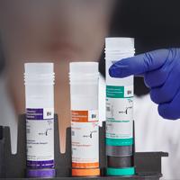
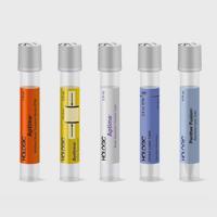
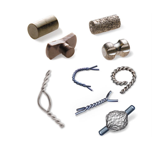
A Comprehensive Range of Compatible Breast Biopsy Markers
The Hologic range of biopsy site markers for use with the Brevera Breast Biopsy Device come in multiple shapes, gauges and lengths. All markers come with blunt tip deployment and an ergonomic and easy to use deployment device included.
Intelligently designed to provide long-term ultrasound visibility, all Tumark markers are non-bioabsorbable, biocompatible permanent markers offering excellent visibility under mammography at deployment and are designed to minimise movement.6
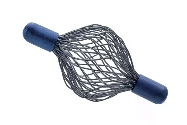
Tumark® Vision
The Tumark Vision Biopsy Site Marker is for use with the Brevera Breast Biopsy Device, and available in both petite and standard size. The unique 3D structure anchors firmly in the tissue, with a mesh structure made of 48 individual wires. Options available: 18G, 127.5mm or 123.5mm length.
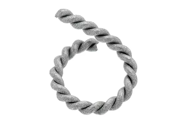
Tumark® Professional Q
The Tumark Professional Q Biopsy Site Marker is for use with the Brevera Breast Biopsy Device, and available in both petite and standard size. The Q-shape becomes firmly anchored in the tissue.5 The slightly open, spiral-shaped design, sandblasted and twisted material designed to provide visibility in ultrasound.6 Options available: 18G, 129.5mm or 125.5mm length.
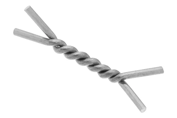
Tumark® Professional X
The Tumark Biopsy Site Marker is for use with the Brevera Breast Biopsy Device, and available in both petit and standard size. Lateral anchor “feet” provide secure anchoring in the tissue5 and the marker is twisted shape with angled ends. Options available: 18G, 127.5mm or 123.5mm length.
SecurMark® Mini Cork
The SecurMark Breast Biopsy Marker is for use with the Brevera Breast Biopsy Device, and available in two different gauge sizes. The marker consists of two pieces, a permanent marker and a bioabsorbable suture-like netting. Options available: 9G or 12G, 130mm length.
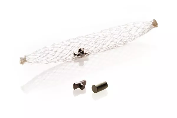
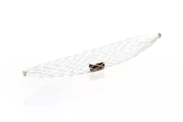
SecurMark® Top Hat
The SecurMark Breast Biopsy Marker is for use with the Brevera Breast Biopsy Device, and available in two different gauge and length sizes. The marker consists of two pieces, a permanent marker and a bioabsorbable suture-like netting. Options available: 9G or 12G, 130mm length.
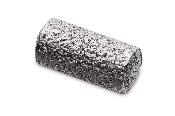
TriMark® Cork
The TriMark Biopsy Site Marker cork shape is for use with the Brevera Breast Biopsy Device, and available in two different gauge sizes. Options available: 9G or 12G, 130mm length.
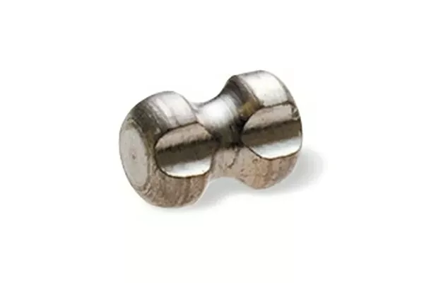
TriMark® Hourglass
The TriMark Biopsy Site Marker hourglass shape is for use with the Brevera Breast Biopsy Device, and available in two different gauge sizes. Options available: 9G or 12G, 130mm length.
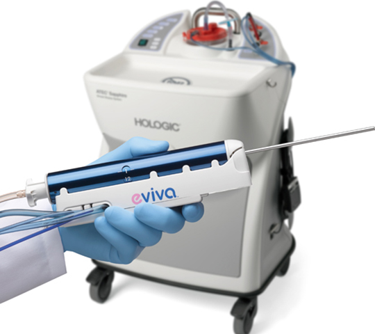
Working with Brevera
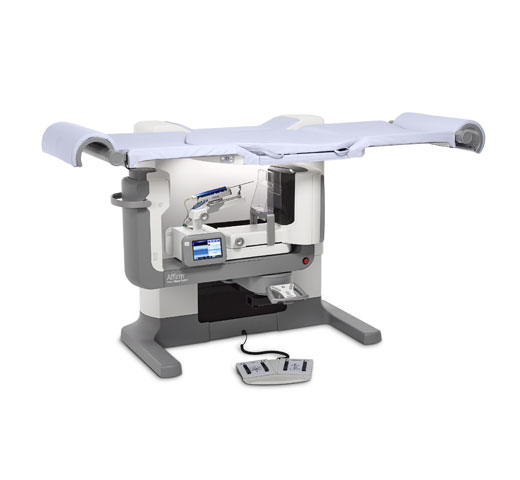
The Ultimate Combination
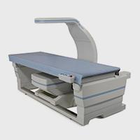
Over 90% of patients agreed that their biopsy procedure was faster and more comfortable than expected when using the Brevera Breast Biopsy System in combination with the Affirm® Prone Breast Biopsy System.10
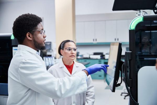
Evidence. Insight. Collaboration.
Our education portal improves patient care through excellence in education, communication of clinical and scientific evidence, and partnerships with the healthcare community.
Insights


- Hologic Data on File: VAR-05326, Rev 2
- Hologic Data on File: MISC-07678-EUR-EN, Rev 1
- Hologic Data on File: MISC-07662-EUR-EN, Rev 1
- Hologic Data on File: DHM-06042, Rev 1
- Hologic Data on File: MISC-07876, Rev 1, attachment 2
- Tumark® Marker Data Collection Study, 2017, DHM-06169, Rev 1, 3 clinicians at 3 hospitals for 90 marker placements, 2017
- Hologic Data on File: VER-07555, Rev 001
- Yen P, Dumas S, Albert A, et al. Post-Vacuum-Assisted Stereotactic Core Biopsy Clip Displacement: A Comparison Between Commercially Available Clips and Surgical Clip. Can Assoc Radiol J. 2018 Feb;69(1):10-15. doi: 10.1016/j.carj.2017.08.004. PMID: 29458952. Devices show reduced displacement when compared to traditional metal surgical clips (28% and 27% vs 38% displacement, p = 0.001 and p = 0.0001). Visibility is dependent on surrounding tissue, experience may vary.
- Pinkney DM, Shah BA. Prospective Comparative Study to Evaluate the Sonographic Visibility of Five Commercially Available Breast Biopsy Markers. Journal of Diagnostic Medical Sonography. 2013;29(4):151-158. doi:10.1177/8756479313486962. *45.4 % of respondence rated SecurMark high sonographic visibility after 6-weeks post procedure, 4x more than the Gel Mark UltraCor™
- Hologic data on file. CSR-00127, Rev 1
Documents
Safety Data Sheets
Package Inserts
2797
0482
Hologic BV, Da Vincilaan 5, 1930 Zaventem, Belgium.
Notified Body number wherever applicable
EC Representative Information wherever applicable