


Future-Proofing Pathology Laboratories

Pathology laboratories across the globe are facing challenges with declining pathologist numbers, the increasing age of the workforce, and greater workload volume and complexity.1
These are expected to increase over the coming years especially in the oncology area with the increasing number of samples that will need analysing. To address these challenges, laboratories are exploring different solutions such as digital pathology, but can it deliver?
Digital pathology includes the acquisition, management, sharing and interpretation of pathology information, including slides and data, in a digital environment.2 Digital slides are created when glass slides are captured with a scanning device, to provide a high-resolution image that can be viewed on a computer screen or mobile device. Its benefits include:2
- Improved laboratory workflow integration and connectivity, which increases the flexibility and efficiency of the workforce.
- Increased ability to share slides and more.
- Rapid referral of cases between organisations or across pathology networks.
Digital tools for supporting slide review, case sharing and digital archiving are now well established in routine diagnostic workflows across many laboratories.3,4 Initially focused on routine histology, their adoption is expanding into more complex cytology and histopathology workflows.
Expanding Role
Digital pathology is now commonly described in pathology labs as being applicable across a broader range of scenarios and working practices, such as remote consultation, second opinions, and participation in multidisciplinary team meetings.
Educational discussions often describe practical considerations when extending use beyond routine applications, including how laboratories may address heterogeneous or fragmented digital infrastructures and varying levels of sample tracking equipment and image management systems.
At the same time, laboratories encounter technical limitations when handling more demanding specimens, particularly where tissue thickness, folding or three-dimensional cellular structure place greater demands on image capture and review.5 These operational and technical constraints are often discussed in the context of exploring different approaches that may support consistent image quality as the scope of digital pathology continues to expand.
The Need for Clarity
Consistently clear digital images are essential for confident interpretation, particularly when reviewing complex cytology and histology specimens.6 Conventional whole-slide imaging is generally described as capturing a single focal plane. This approach may be suited to thin, uniform sections, and may present limitations in clarity when tissue thickness varies or when three-dimensional structures are present.
Some digital pathology systems use z-stacking for these samples, acquiring multiple images at different focal depths and combining them to improve visualisation across uneven areas of a specimen.5 This approach is often described in pathology lab in general, as enhancing focus in selected regions, while also increasing scan time and data volume, and it may be associated with variable image quality across the region of interest for entire tissue on slide.
The unified Digital Pathology Solution powered by advanced volumetric imaging enables digitisation and review of cytology and histology in one system.7,8 The data from the studies conducted on the GeniusTM Digital Diagnostics System demonstrate that the Genius Digital Diagnostics System provides images that may be reliably reviewed for diagnostic evaluation of cytology and surgical pathology specimens.8
Scanning Complex Samples
The study data show the Sample Detect scan profile reliably identifies the areas of the slide that contain specimen, and the Genius Digital Diagnostics System reliably creates digital images of pathology microscope slides that are suitable for review.8 Therefore, pathology specimens may be reliably reviewed for diagnostic evaluation using this system.8
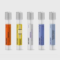
Folds and wrinkles
Tissue sections with folds or wrinkles are generally not perfectly flat, which can make it difficult to maintain consistent image quality across the entire section.
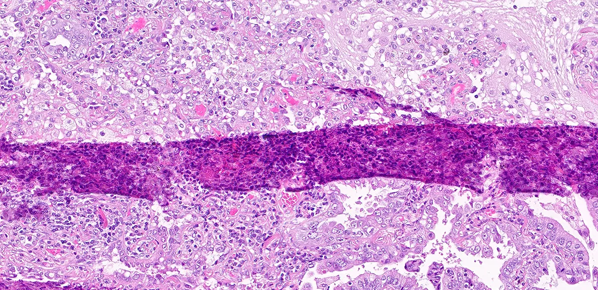
Figure 1: Example volumetric scan of a 20× tissue section obtained using the Genius Digital Diagnostics System, illustrating a high density of folds and surface unevenness (Snapshot of Image taken from Genius Review Station for educational purposes only).
Fatty, thick or fragmented tissue
Fatty tissue specimens in pathology can require an extra care during lab processing due to their translucency and variable thickness, which may make consistent focus more difficult to achieve.
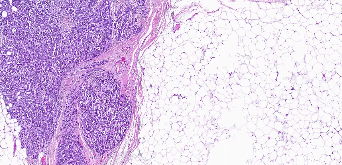
Figure 2: Example fatty tissue section imaged with volumetric scanning on the Genius Digital Diagnostics System (Snapshot of Image taken from Genius Review Station for educational purposes only).
Cytology and histopathology specimens
Three-dimensional cell clusters and histopathology specimens often contain mixed cell populations distributed across multiple focal depths.
Figure 3: Example of Genius Review station showing diverse specimen types (Gyn, NGYN and Histology) (Image for educational purposes only).
Volumetric imaging enables effective digital scanning for both cytology and histology, with 14 focal planes captured in a single pass and merged to an exceptionally clear, 2-dimensional image.7,8
Real-World Experience
Quote from Professor Paul van Diest,
“The ability to image and review more types of samples within a single system will help us think beyond traditional boundaries and provide greater precision and efficiency in our work.”
Quote from Dr. Ingibjörg Guðmundsdóttir,
“With just one other cytopathologist and a single technician available, I realised that the Genius Digital Diagnostics System was the only viable way to keep the screening service running.”
A Bright Future for Digital Pathology
As digital pathology continues to evolve, laboratories are increasingly described as looking beyond routine digitisation towards approaches that can accommodate a broader range of specimens and workflows. In this context, moving from digitising individual processes to considering entire diagnostic pathways is often associated with a greater focus on managing cytology and histology workflows within a single platform.
Find out more here.
For more insight articles, visit the Hologic Innovation Exchange.
2797
Hologic BV, DA Vincilaan 5,
1930 Zaventem, Belgium
Notified Body number wherever applicable
Walsh E, Orsi NM. The current troubled state of the global pathology workforce: a concise review. Diagn Pathol. 2024 Dec 21;19(1):163.
Digital Pathology. https://www.rcpath.org/profession/digital-pathology.html last accessed 30 April 2026.
Bessen JL et al. Perspectives on Reducing Barriers to the Adoption of Digital and Computational Pathology Technology by Clinical Labs. Diagnostics. 2025;15(7):794. doi:10.3390/diagnostics15070794.
Pantanowitz L et al. Twenty Years of Digital Pathology: An Overview of the Road Travelled, What is on the Horizon, and the Emergence of Vendor-Neutral Archives. Journal of Pathology Informatics. 2018;9(1).
Wildrick M. Crystal Clear: Why Image Quality Matters. The Pathologist. (2025) Accessed 22 January 2026. https://www.thepathologist.com/issues/2025/articles/september/crystal-clear-why-image-quality-matters/.
Ahuja S, Zaheer S. Advancements in Pathology: Digital Transformation, Precision Medicine, and Beyond. Journal of Pathology Informatics. 2025;100408.
Genius™ Digital Diagnostics | Digital Pathology | Hologic® UK, https://www.hologic.co.uk/en-gb/products/genius-digital-diagnostics-histology Website last accessed on 31st March 2026.
Genius Digital Diagnostics System Whole Slide Imaging Instructions for use. AW- 32577-002_Rev 001, Hologic Inc., 2025.